Contents
Overview
The concept of visualizing chromosomes as a distinct set began to coalesce in the late 19th and early 20th centuries, building upon earlier observations of cellular division. Early cytologists like Walther Flemming described chromosome behavior during mitosis in the 1880s, laying the groundwork for understanding these structures. However, the systematic organization and interpretation of chromosomes into what we now recognize as a karyotype gained momentum with advancements in microscopy and staining techniques. The crucial step of fixing cells and spreading chromosomes was refined by researchers like Theophilus Pennell King and Leonard Coleman Strong in the 1920s and 1930s. A pivotal moment arrived in 1956 when Joe Hin Tjio and Albert Levan definitively established the human diploid chromosome number as 46, correcting decades of misconception and providing a standardized basis for human karyotyping. This discovery, published in Hereditas, revolutionized human genetics and paved the way for clinical applications.
⚙️ How It Works
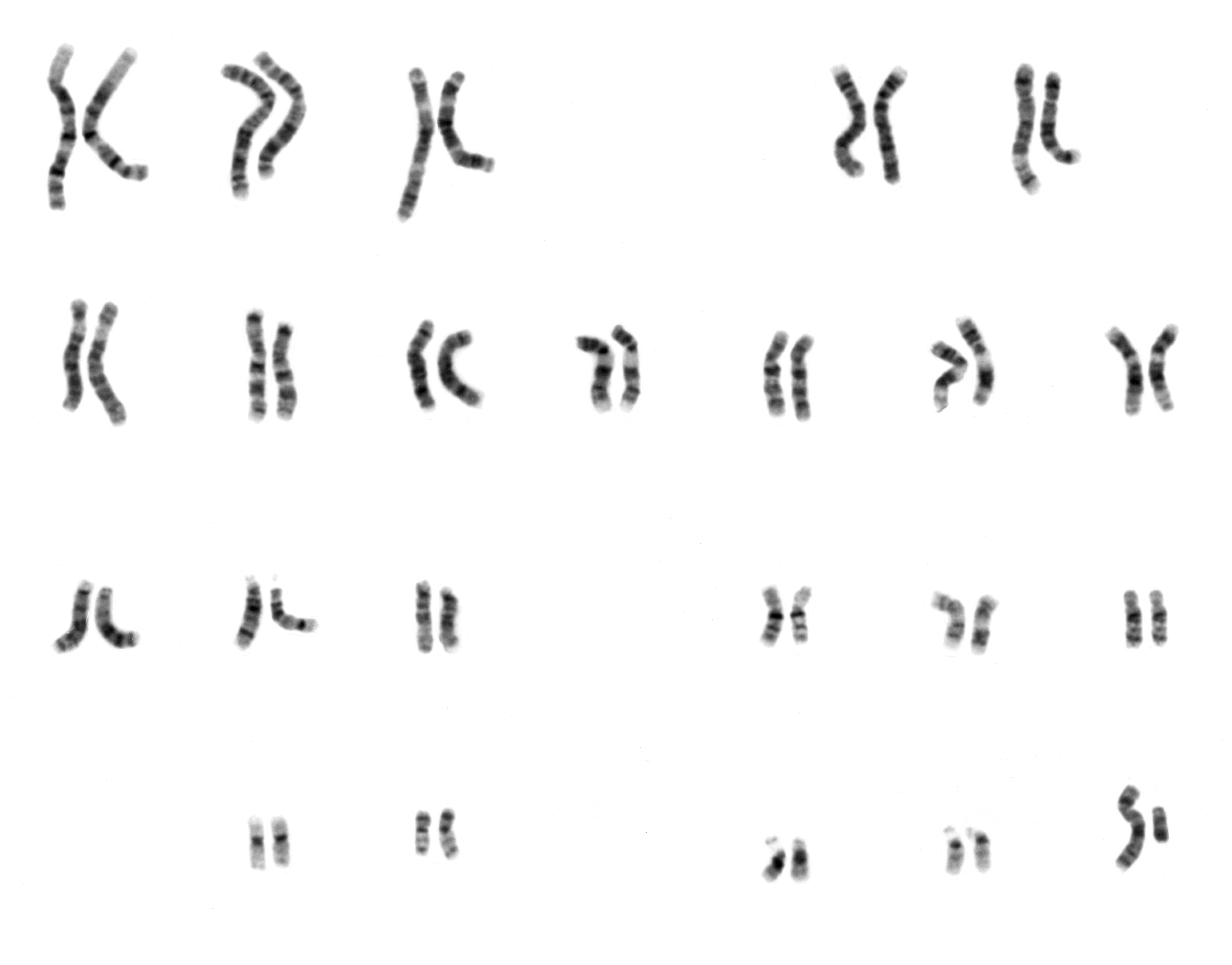
Karyotyping involves several key steps, beginning with obtaining a cell sample, often from blood, amniotic fluid, or tissue biopsies. These cells are cultured in a laboratory to stimulate cell division. During metaphase, when chromosomes are most condensed and visible, a chemical agent like colchicine is added to halt the cell cycle. The cells are then treated to swell and burst, dispersing the chromosomes. These spread chromosomes are stained, often with Giemsa stain, which produces characteristic banding patterns (e.g., G-banding) that help identify individual chromosomes and their regions. High-resolution images are captured using microscopy and digital photography, and then the chromosomes are digitally or manually arranged into homologous pairs, ordered by size and centromere position, creating the karyogram. This organized display allows for the detection of aneuploidies (abnormal chromosome numbers) and structural rearrangements like translocations or deletions.
📊 Key Facts & Numbers
The standard human karyotype consists of 46 chromosomes, arranged as 23 pairs: 22 pairs of autosomes and one pair of sex chromosomes. Females have XX sex chromosomes, while males have XY sex chromosomes. Deviations from this number are significant; for instance, Down syndrome is caused by trisomy 21, meaning an individual has three copies of chromosome 21 instead of two, totaling 47 chromosomes. Turner syndrome (45, X) and Klinefelter syndrome (47, XXY) are common sex chromosome aneuploidies. The resolution of karyotyping can detect chromosomal abnormalities as small as 5-10 megabases (Mb). Globally, approximately 1 in 160 live births worldwide are affected by a major chromosomal abnormality detectable by karyotyping. The cost of a standard karyotype analysis can range from $200 to $1000 USD, depending on the laboratory and complexity.
👥 Key People & Organizations
Key figures in the development of karyotyping include Joe Hin Tjio, who, along with Albert Levan, correctly identified the human chromosome number in 1956. Jérôme Lejeune famously used karyotyping in 1959 to identify the extra chromosome 21 responsible for Down syndrome. Organizations like the American Society of Human Genetics and the International Human Genome Sequencing Consortium have been instrumental in advancing genetic research, including the refinement of cytogenetic techniques. Major diagnostic laboratories and research institutions worldwide, such as the Mayo Clinic and Broad Institute, routinely perform and develop improved karyotyping methods.
🌍 Cultural Impact & Influence
The ability to visualize an organism's entire chromosomal set has profoundly impacted our understanding of genetics and inheritance. Karyotypes have become a visual shorthand for genetic health, appearing in medical textbooks, patient education materials, and even popular science discussions about genetic predispositions. The discovery of specific chromosomal abnormalities linked to diseases like chronic myeloid leukemia (the Philadelphia chromosome, a translocation between chromosomes 9 and 22) demonstrated the diagnostic power of karyotyping. This visual representation of genetic material has fostered a deeper public awareness of genetic diversity and the biological basis of certain conditions, influencing societal perceptions of disability and genetic screening.
⚡ Current State & Latest Developments
While traditional karyotyping remains a vital diagnostic tool, its resolution is being surpassed by newer technologies. Whole-genome sequencing and chromosomal microarray analysis (CMA) can detect smaller deletions and duplications (copy number variations) that are often invisible to standard karyotyping. However, karyotyping's strength lies in its ability to detect balanced translocations and complex rearrangements, which CMA might miss. Current developments focus on improving the speed and accuracy of karyotyping, integrating it with molecular techniques, and applying artificial intelligence for automated analysis of karyograms. The ongoing debate centers on when to use karyotyping versus CMA or sequencing for specific diagnostic scenarios.
🤔 Controversies & Debates
The primary controversy surrounding karyotyping lies in its resolution limitations compared to newer molecular techniques. While it can identify aneuploidies and large structural rearrangements, it often fails to detect smaller, submicroscopic deletions or duplications that can cause significant genetic disorders, a limitation addressed by chromosomal microarray analysis (CMA). Conversely, CMA may miss balanced translocations that can lead to reproductive issues. Another debate concerns the interpretation of variants of unknown significance (VUS) identified during karyotyping or CMA, which can cause anxiety for patients and families. Ethical considerations also arise regarding the use of karyotyping in prenatal diagnosis and the implications of identifying genetic predispositions.
🔮 Future Outlook & Predictions
The future of karyotyping likely involves its integration with advanced molecular technologies rather than outright replacement. Next-generation sequencing (NGS) technologies are becoming increasingly cost-effective and capable of providing comprehensive genomic information, including chromosomal abnormalities. However, karyotyping's ability to visualize the entire chromosome set and detect balanced rearrangements will likely ensure its continued relevance, particularly in specific clinical contexts like cancer cytogenetics and reproductive genetics. Research is ongoing to develop higher-resolution banding techniques and AI-driven analysis platforms that could enhance the diagnostic yield of traditional karyotyping, potentially making it more competitive with molecular methods for certain applications.
💡 Practical Applications
Karyotyping is indispensable in clinical genetics for diagnosing a wide range of conditions. It's routinely used in prenatal diagnosis to screen for chromosomal abnormalities like Down syndrome, Edwards syndrome, and Patau syndrome in fetuses. Postnatally, it aids in diagnosing developmental delays, intellectual disabilities, congenital anomalies, and infertility. In oncology, karyotyping is critical for classifying and monitoring cancers, identifying specific chromosomal translocations that drive tumor growth, such as the Philadelphia chromosome in CML. It also plays a role in assessing the genetic stability of cell lines used in research and biotechnology.
Key Facts
- Category
- science
- Type
- topic